X-rays are a form of electromagnetic radiation with a wavelength shorter than visible light, making them invisible to the human eye. X-ray technology is used in medical imaging, radiography, radiation therapy, and other diagnostic imaging techniques. How do x-rays work, and what are the principles behind their use? In this article, we will explore the production of x-rays, how they interact with matter, and their applications in medicine.
I. X-Ray Production
X-rays are produced when high-energy electrons collide with a material, typically a metal target. This process is called bremsstrahlung radiation, which translates to “braking radiation” in German. When an electron is slowed down or stopped by the target’s atomic nucleus, it loses energy and emits an x-ray photon. The energy of the x-ray photon is determined by the energy of the original electron and the atomic structure of the target material.
II. Interactions with Matter
X-rays interact with matter in different ways, depending on their energy and the nature of the material. When x-rays pass through a material, they may be absorbed, scattered, or transmitted. The probability of each interaction occurring depends on the energy of the x-ray and the atomic number of the material. Higher atomic numbers result in more absorption and scattering.
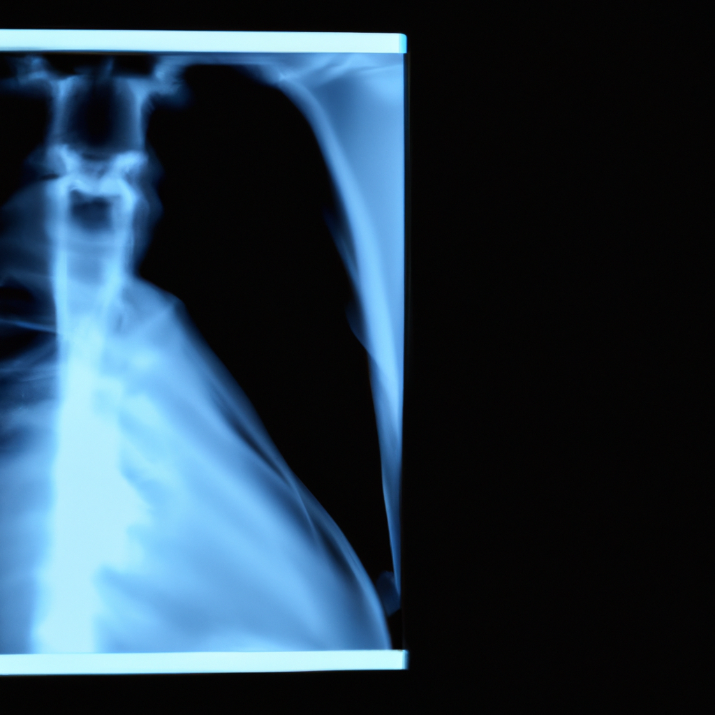
III. Medical Applications
X-rays have numerous medical applications, primarily in imaging and radiation therapy. X-ray imaging uses the differential absorption of x-rays by various tissues to generate an image. This technique is particularly useful for visualizing bones, which have a high atomic number and absorb x-rays more than soft tissue. Radiography is commonly used for diagnosing fractures, dislocations, and other skeletal abnormalities.
IV. Diagnostic Imaging Techniques
Other diagnostic imaging techniques that use x-rays include computed tomography (CT) and mammography. CT imaging uses x-rays to construct a 3D image of the body, allowing doctors to visualize internal structures in detail. Mammography is a specialized form of x-ray imaging used to detect breast cancer.
V. Radiation Therapy
X-rays are also used in radiation therapy to treat cancer. In this application, high-energy x-rays are targeted at cancerous tissue to destroy or damage the cancer cells. The goal is to deliver a dose of ionizing radiation that is high enough to kill the cancer cells but not so high that it damages healthy tissue.
VI. X-Ray Machine
X-ray machines consist of three main components: the x-ray tube, the power supply, and the control panel. The x-ray tube contains the metal target and produces the x-rays. The power supply provides the high voltage needed to accelerate the electrons and generate the x-rays. The control panel allows the operator to adjust the exposure settings and activate the x-ray tube.
VII. Ionizing Radiation
One potential drawback of x-rays is that they are a form of ionizing radiation, which means they have enough energy to ionize atoms and molecules. This can damage DNA and other cellular structures, potentially leading to cancer or other health problems. To minimize the risks of radiation exposure, x-ray technicians and other medical professionals must follow strict safety protocols and limit unnecessary exposure.
In conclusion, x-rays are a vital tool in medical imaging and radiation therapy. By understanding the principles of x-ray production and interaction with matter, we can appreciate their applications in medicine and the potential risks associated with their use. With advances in technology, x-ray imaging has become safer and more precise, allowing doctors to diagnose and treat a wide range of health conditions.