When it comes to medical imaging, magnetic resonance imaging (MRI) is a widely used technology that allows doctors to get a detailed look inside the human body. MRI scans are used to identify a wide range of conditions, from injuries and tumors to neurological disorders and cardiovascular disease. But how does an MRI scanner work? In this article, we’ll explore the physics behind MRI technology, how an MRI machine creates images, and what happens during an MRI procedure.
MRI Physics
Magnetic resonance imaging is based on the physical properties of atoms and molecules. Every atom has a nucleus, which is made up of protons and neutrons. Protons have a positive charge, while neutrons have no charge. When a magnetic field is applied to a substance, the protons align themselves with the field.
When the magnetic field is switched off, the protons return to their original orientation. This process releases energy, which can be detected by a receiver coil. By analyzing the signals from the receiver coil, an MRI machine can create images of the body.
MRI Anatomy
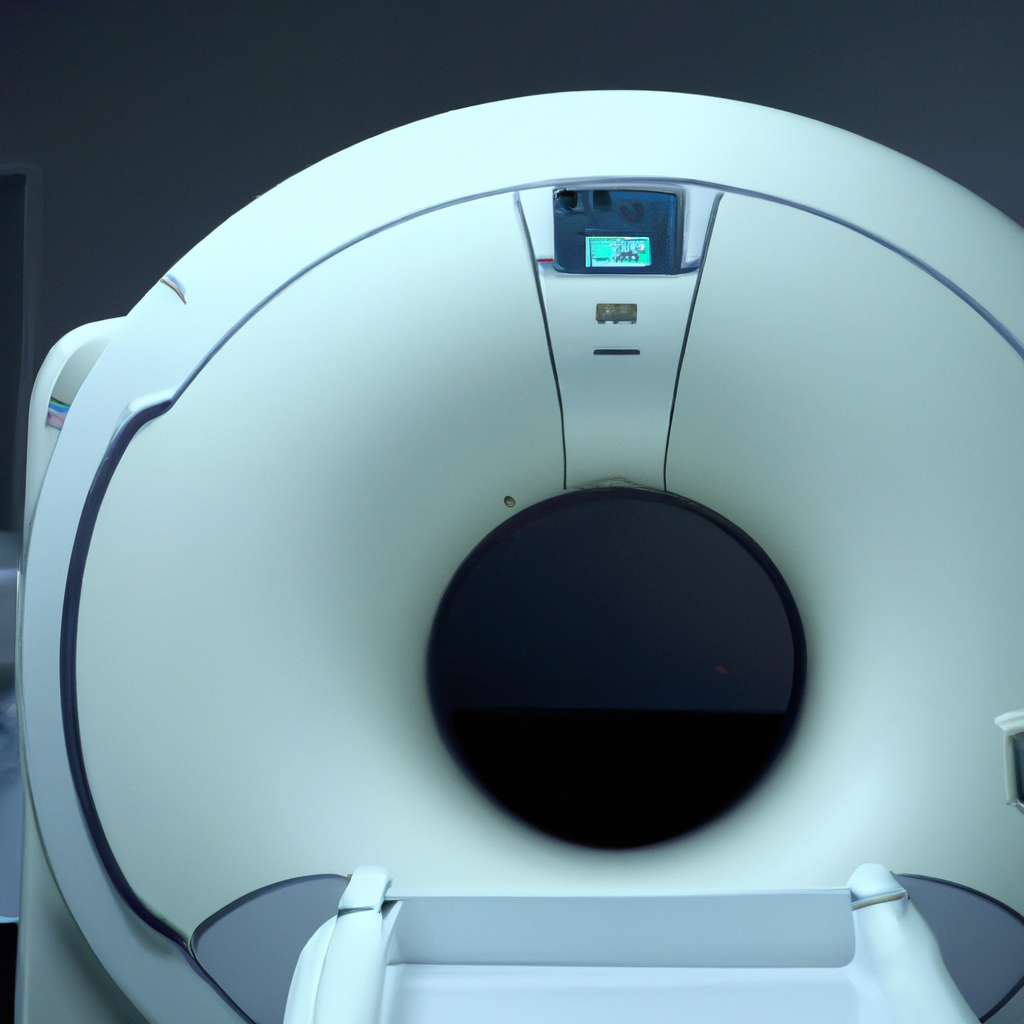
Before we dive into the specifics of how an MRI scanner works, let’s take a quick look at the anatomy of an MRI machine. An MRI scanner consists of a large cylindrical magnet, a radiofrequency transmitter and receiver, and a computer that controls the entire process.
The patient lies on a table that slides into the magnet, which creates a strong magnetic field. The radiofrequency transmitter sends a series of pulses into the patient’s body, which cause the protons in the body to emit energy. The receiver coil detects this energy and sends it to the computer, which uses the data to create an image of the body.
How Does MRI Work?
Now that we have a basic understanding of the physics and anatomy of an MRI machine, let’s explore how an MRI scan actually works.
Step 1: Preparation
Before the scan begins, the patient is asked to remove any metal objects, such as jewelry or watches, as these can interfere with the magnetic field. The patient then lies on the table, which slides into the magnet.
Step 2: Alignment
Once the patient is in the magnet, the MRI technician uses lasers to align the patient’s body with the scanner. This ensures that the images are as clear and accurate as possible.
Step 3: Radiofrequency Pulses
The radiofrequency transmitter sends a series of pulses into the patient’s body, which cause the protons in the body to emit energy.
Step 4: Signal Detection
The receiver coil detects the energy emitted by the protons, and sends the data to the computer.
Step 5: Image Creation
The computer uses the data to create a detailed image of the patient’s body. The image can be viewed in real-time, allowing the technician to make any necessary adjustments.
Step 6: Completion
Once the scan is complete, the patient can leave the machine. The images are saved and sent to a radiologist, who will interpret the results and provide a report to the patient’s doctor.
Benefits of MRI Imaging
MRI imaging offers a number of benefits over other types of medical imaging. MRI scans are non-invasive and do not use ionizing radiation, making them safer than X-rays and CT scans. MRI scans can also provide more detailed images of soft tissue, making them a valuable tool for diagnosing conditions such as tumors and neurological disorders.
Conclusion
In summary, magnetic resonance imaging (MRI) is a powerful medical imaging technology that allows doctors to get a detailed look inside the human body. MRI machines use a combination of magnets, radiofrequency pulses, and sophisticated computer algorithms to create images of the body. By understanding the physics and anatomy of MRI technology, patients can have a better understanding of what happens during an MRI procedure and the benefits of MRI imaging.