Ultrasound technology has revolutionized the field of medical imaging and diagnostic imaging by making it possible to see inside the body without invasive procedures. Ultrasound machines have become a vital tool in medical settings, providing a non-invasive way to visualize internal organs, tissues, and structures. In this article, we will explore how an ultrasound machine works and the science behind this technology.
What is an Ultrasound Machine?
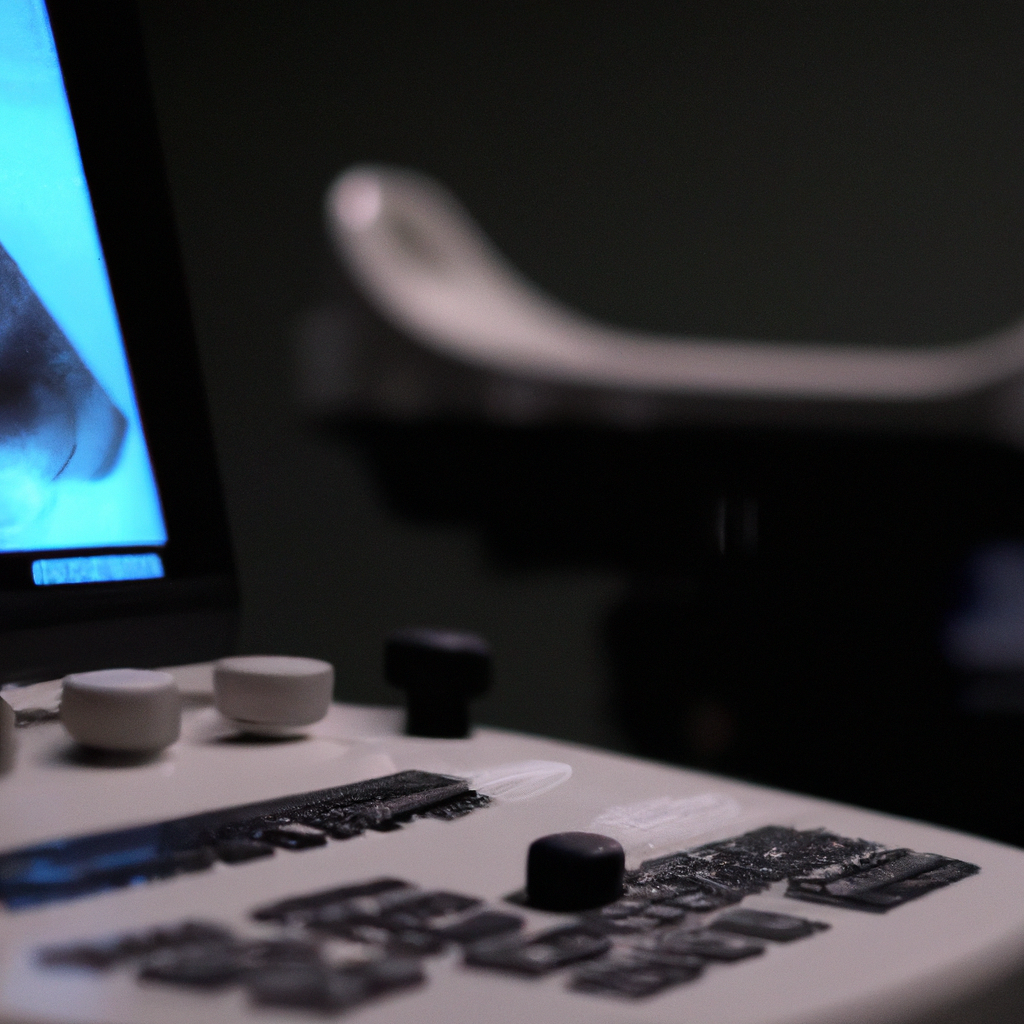
An ultrasound machine is a medical device that uses ultrasound waves to create images of internal organs and tissues. It is also known as sonography or ultrasound imaging. The machine consists of an ultrasound probe, a computer, and a display screen. The probe emits high-frequency sound waves that penetrate the body and bounce back to the probe. The computer then processes these waves to create images that are displayed on the screen.
How does an Ultrasound Machine Work?
An ultrasound machine works by using ultrasound waves to create images of internal structures in the body. These waves are high-frequency sound waves that cannot be heard by humans. The ultrasound probe emits these waves, which penetrate the body and bounce back to the probe. The probe then detects these waves and sends them to the computer for processing. The computer uses the information from the waves to create images of internal organs and tissues.
The ultrasound waves used in medical imaging are much higher in frequency than the sound waves we hear in everyday life. These waves typically range from 1 to 18 megahertz (MHz). The higher the frequency, the more detailed the image will be. However, higher frequency waves have less penetration, so the depth of penetration needs to be balanced with the resolution needed for the specific examination.
The ultrasound probe is the main component of the machine that emits and receives ultrasound waves. The probe contains a piezoelectric crystal that vibrates when an electric current is applied to it. This vibration creates the ultrasound waves that penetrate the body. The same crystal also detects the waves that bounce back to the probe and converts them into an electric signal that is sent to the computer for processing.
The computer receives the signals from the ultrasound probe and processes them to create images. The computer uses advanced algorithms to analyze the signals and create images that are displayed on the screen. The images are created in real-time, which means that the operator can see the internal structures of the body as they move and function in real-time.
Ultrasound imaging is a safe and non-invasive procedure that does not use ionizing radiation, unlike other medical imaging techniques such as X-rays and CT scans. It is also relatively inexpensive compared to other imaging techniques, making it accessible to a wide range of patients.
Applications of Ultrasound Imaging
Ultrasound imaging is used in a wide range of medical applications, including obstetrics and gynecology, cardiology, urology, and gastroenterology. It is commonly used to visualize the fetus during pregnancy, diagnose and monitor heart conditions, detect and monitor the progression of cancer, and diagnose and treat conditions of the urinary tract and digestive system.
Advancements in Ultrasound Technology
Ultrasound technology has come a long way in recent years, with advancements in technology making it possible to create more detailed and accurate images. Newer ultrasound machines use 3D and 4D imaging, which provide a more comprehensive view of the internal structures of the body. These machines also use advanced software that can automatically detect and measure structures in the body, making diagnosis and treatment more accurate and efficient.
Conclusion
In conclusion, ultrasound technology has revolutionized the field of medical imaging and diagnostic imaging by providing a non-invasive way to visualize internal organs, tissues, and structures. Ultrasound machines work by using high-frequency sound waves to create images of internal structures in the body. The ultrasound probe emits these waves, which bounce back to the probe and are processed by a computer to create images that are displayed on a screen. Ultrasound imaging is a safe and non-invasive procedure that is used in a wide range of medical applications, and advancements in technology are making it possible to create even more detailed and accurate images.